
From Skin Cells to Functional Muscle: How AI-Designed Proteins and Mantarray Are Rewriting Muscle Regeneration
When AI Meets Cell Biology, Muscle Regeneration Gets a Whole New Playbook 💪
What if the cells needed to repair aging or diseased muscle were hiding in plain sight — in your own skin?
That's not a hypothetical. A landmark new study from the University of Washington, published on bioRxiv in April 2026, shows that AI-designed proteins can be used to reprogram ordinary skin cells directly into functional human muscle tissue, and that Curi Bio's Mantarray™ platform was the critical tool used to prove those tissues actually function.
The study, "Designed Minibinders Rewire Receptor Signaling to Enable Functional Human Myogenic Reprogramming," comes from the labs of Dr. Hannele Ruohola-Baker, Dr. David L. Mack, and Nobel laureate Dr. David Baker at the University of Washington's Institute for Stem Cell and Regenerative Medicine (ISCRM). The Mantarray experiments in this study were completed by our very own Phillip Barrett, a Curi Bio scientist and UW postdoctoral researcher with the Mack Lab.
Why Muscle Regeneration Matters More Than Ever
Most of us don't think much about skeletal muscle until it starts to fail. But here's the reality: muscle strength begins to decline as early as our 30s. By the time many people reach their 70s and 80s, the loss is significant enough to affect independence, quality of life, and survival. This condition called sarcopenia, is one of the most prevalent and underappreciated health burdens in aging populations worldwide.
It's not just aging. Severe trauma, surgical injury, and genetic diseases like Duchenne muscular dystrophy (DMD) can devastate muscle at any age. And more recently, the widespread use of GLP-1 receptor agonist medications (semaglutide, tirzepatide, etc.) has brought fresh attention to the problem: while these drugs are transformative for weight loss, they can also cause significant loss of lean body mass, potentially accelerating sarcopenia in vulnerable patients.
The field has long dreamed of a way to regenerate functional human muscle, not just grow cells in a dish, but create tissue that actually contracts, generates force, and behaves like the real thing.
The Big Idea: Reprogramming, Not Rebuilding from Scratch
The conventional approach to generating new muscle cells in the lab involves reprogramming cells all the way back to a pluripotent stem cell state, then coaxing them forward again, a slow, complex process. An alternative vision, called direct reprogramming (or transdifferentiation), skips the detour entirely: you take an adult cell, like a skin fibroblast, and convert it directly into a muscle cell.
The concept has been around since the 1980s, when researchers discovered that a single gene, MyoD, could flip fibroblasts toward a muscle identity. But decades of follow-up work revealed a harsh truth: getting cells to start expressing MyoD is the easy part. Getting them to actually become functional, mature muscle cells is much harder. Competing molecular signals, pro-fibrotic pathways, inflammatory programs, stiff extracellular scaffolding, all conspire to keep cells anchored to their original identity.
The problem isn't the transcription factor. The problem is the signaling environment the cell is living in.
Enter the AI-Designed Minibinders
Over the past several years, the Baker Lab at UW has pioneered the use of AI-based protein design tools, including RFDiffusion and BindCraft, to create entirely new proteins from scratch. These aren't modified natural proteins; they're de novo designed molecules, built computationally to bind specific receptor targets with exquisite precision.
The researchers hypothesized that if the primary obstacles to muscle conversion are rooted in the extracellular signaling environment, then the very proteins anchoring fibroblasts to their original identity could be bypassed using computationally engineered modulators to systematically dismantle these developmental checkpoints. They screened a library of AI-designed "minibinders" targeting a range of surface receptors and, through systematic testing, identified a cocktail they named C6-DPC (Designed Protein Cocktail 6).
C6-DPC combines three components working in concert:
An activator of FGFR1/2c: a growth factor receptor that promotes muscle-cell identity
An inhibitor of ALK1: a receptor that normally keeps cells in a non-muscle state
An inhibitor of TGFBR2: a receptor that drives the pro-fibrotic program that competes with muscle fate
The result: fibroblast-to-muscle conversion efficiency jumped more than 3-fold compared to standard conditions, and the resulting myotubes were larger, exhibited better fusion, and more metabolically active than controls.
A key bonus finding: inflammation acts as a dominant checkpoint in this process. When the team added a designed inhibitor against the gp130 receptor, a key node in inflammatory signaling, conversion efficiency climbed even higher. The message is clear: to make muscle, you have to not only turn on the right signals, but also quiet the inflammatory noise that keeps the door locked.
From 2D to 3D: Proving It in Human Engineered Muscle Tissue
Cultivating cells on a plate is just the beginning. Validating that they can assemble into functional 3D tissues capable of generating real force is the bigger hurdle and the team used the Mantarray platform to redefine the experimental landscape.
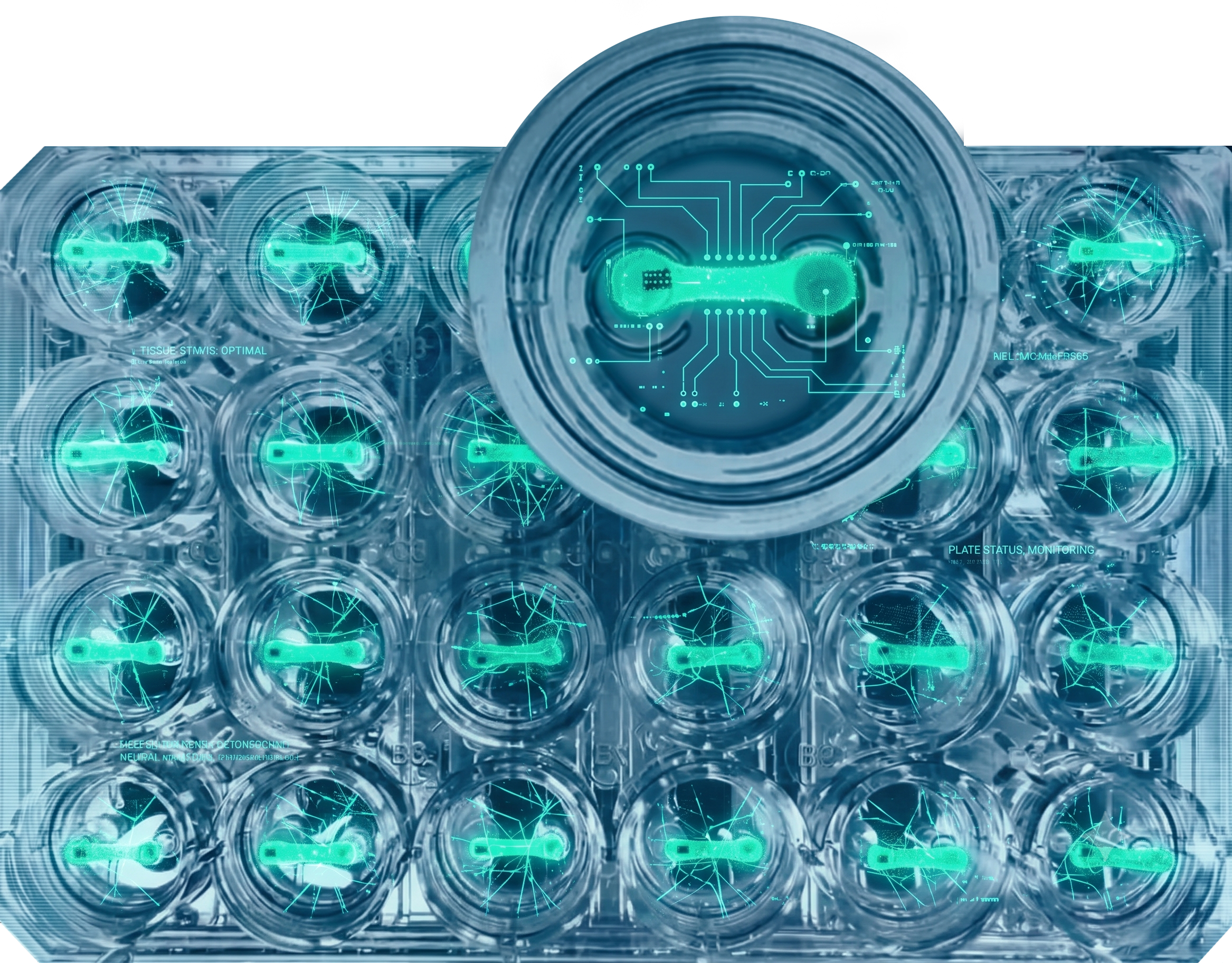
Curi Bio's Mantarray platform was used to generate and evaluate 3D engineered muscle tissues (EMTs) from iPSC-derived myoblasts in the presence or absence of C6-DPC treatment. The Mantarray system uses a magnetometric approach to measure real-time contractile force in 3D tissues, giving researchers a direct, quantitative window into whether their cells are behaving like real muscle.
The functional results were striking:
In healthy, control tissues:
C6-DPC treatment increased specific tetanic force by approximately 1.6x (1.03 → 1.69 mN/mm²)
Specific twitch force also increased by ~1.6x
Tissues showed faster relaxation kinetics, a hallmark of mature skeletal muscle
In dystrophin-deficient DMD tissues:
Specific tetanic force increased by a profound ~3x (0.70 → 2.10 mN/mm²)
Specific twitch force increased by an astonishing ~4.5x
Relaxation kinetics improved significantly
Why This Matters: A New Framework for the Field
This paper shifts the conversation in at least three important ways.
Figure 1: Specific tetanic and twitch forces in WT and DMD engineered muscle tissues demonstrate that C6-DPC treatment significantly increased contractile force over controls in both genotypes, with the largest gains in DMD tissues.
First, it establishes that AI-designed proteins can serve as precision tools for controlling cell fate. Unlike small molecules, which tend to be blunt instruments affecting many pathways at once, these minibinders are engineered to hit specific receptor isoforms. The C6-DPC cocktail doesn't just nudge cells toward muscle, it systematically dismantles the barriers while simultaneously activating the right programs. This is a level of molecular control the field hasn't had before.
Second, it reframes the role of inflammation in muscle regeneration. The discovery that inflammatory signaling can completely override pro-myogenic cues, and that a designed inhibitor can lift this ceiling, has broad implications. In aging tissue, after injury, and in disease states like DMD, chronic inflammation is a constant challenge. Knowing how to specifically target these signals without broadly suppressing the immune system opens new therapeutic avenues.
Third, it shows functional rescue in DMD models, even without fixing the gene. The DMD community has invested heavily in dystrophin restoration strategies (gene therapies, exon skipping, etc.), but functional recovery often remains incomplete even with partial genetic correction. This study hints that maturation-targeted signaling strategies could be complementary, helping the body make more out of whatever muscle it does have.
Beyond muscle disease, the implications extend to the growing clinical challenge of GLP-1-associated lean mass loss. As millions of patients use semaglutide and similar drugs long-term, preserving and regenerating skeletal muscle becomes a genuine public health imperative. Precision tools like C6-DPC, potentially refined and translated into therapeutically applicable forms, represent a compelling path forward.
A Closer Look at the Tissues
A few things stand out in these images. Dystrophin-deficient (DMD) engineered muscle tissues lack the peripheral dystrophin localization around the myotubes seen in the controls, while the C6-DPC minibinder cocktail produced denser, more organized myofiber architecture in both control and DMD tissues. This structural improvement was reflected in the functional contractility data collected with Mantarray.
The Role of Mantarray: Measuring What Matters
Muscle biology ultimately lives and dies on function. You can stain cells for muscle markers, sequence their transcriptomes, and image their sarcomeres, and that is valuable. But if the tissue doesn't contract, doesn't generate force, doesn't respond to electrical stimulation like real muscle, then all the molecular markers in the world don't tell you whether you've actually achieved your goal.
Mantarray provides exactly this readout: real-time, non-invasive contractile force measurement from living 3D tissues. By enabling the UW team to measure twitch force, tetanic force, and relaxation kinetics in both healthy and DMD engineered tissues, Mantarray transformed this study from a promising cell biology story into a validated functional proof-of-concept.
Curi Bio builds 3D human-relevant tissue platforms to give researchers the tools to ask, and definitively answer, the question that matters most: does it work?
Figure 2: Immunofluorescence cross-sections of WT and DMD EMTs treated with PBS or C6, stained for nuclei (blue), myosin heavy chain (green), and dystrophin (red).
What Comes Next
The researchers point toward future applications including muscle repair after injury, degenerative disease modeling, precision regenerative medicine, and addressing the growing challenge of drug-associated sarcopenia.
For the scientific community, this work provides a new methodological blueprint: use AI to design the proteins, use direct reprogramming to convert the cells, use 3D functional tissue models to validate the outcome.
Learn More About Curi Bio's 3D Tissue Platforms
If your research involves skeletal muscle biology, regenerative medicine, or functional tissue engineering, Curi Bio offers the most advanced 3D human-relevant tissue platforms available today.
👉 Explore Mantarray — the platform powering the next generation of muscle biology research.
👉 View our Skeletal Muscle Models — from 2D cell culture to 3D engineered muscle tissues, built for human-relevant discovery.
👉 Contact our Team — to learn how Curi Bio can support your research program.
Publication: Keshri R, Foreman Z, Barrett P, et al. "Designed Minibinders Rewire Receptor Signaling to Enable Functional Human Myogenic Reprogramming." bioRxiv, April 2026. DOI: https://doi.org/10.64898/2026.04.26.720818