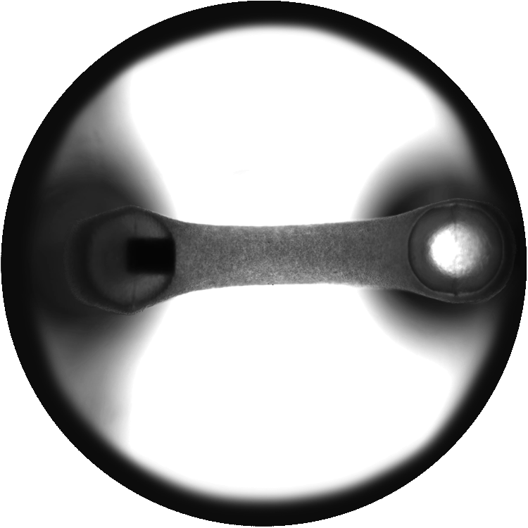

3D Engineered Heart Tissues (EHTs)
3D Engineered Heart Tissues (EHTs) Enable in vitro Models of Cardiac Muscle Contractility
Allows assessment of changes to tissue function in response to disease or drug treatment.
Reliable and reproducible tissue casting across entire plate (over 95% success rate).
Learn more about Mantarray – Curi Bio’s novel 3D Engineered Muscle Tissue platform that uses facile and scalable bioengineering approaches to generate tissues from a variety of cell sources, and a label-free parallel measurement technique:
Curi Bio's 3D EHT Model Allows Measurement of:
- Contractile Force
- Force Frequency
- Rate of Contraction/Systole
- Rate of Relaxation/Diastole
- Time to Max Twitch (Peak)
- Maximal Twitch Amplitude
- Full Width at Half Max
Application Examples
Advance Your Research with 3D Engineered Heart Tissues (EHTs) on Curi Bio’s Mantarray.
Data Analysis
Simultaneous analysis of data from all 24 wells using Curi Bio’s custom data package.
Easily accessible user report showing clinically relevant functional data.
Toxicology/Safety Screening
Tissue responses to drugs can be monitored on short (minutes) or long (days) timescales.
High throughput measurement technique capable of direct contractility assessment across 24 tissues in parallel.
BMS-986094: Graph showing twitch frequency (Hz) over 14 days.
Click Image to Enlarge
Disease Modeling
Development of cardio models for gene therapy and AAV gene editing.
Derivation of an ultra-rare patient specific Duchenne’s Muscular Dystrophy models for personalized gene therapy.
Hypertrophic cardiomyopathy modeling of therapeutic candidates.


Curi Bio's Mantarray Instrument
The Mantarray Platform brings functional data into the earliest stages of preclinical testing of new medicines.
Learn more about how Curi Bio’s Mantarray Platform can benefit your lab:
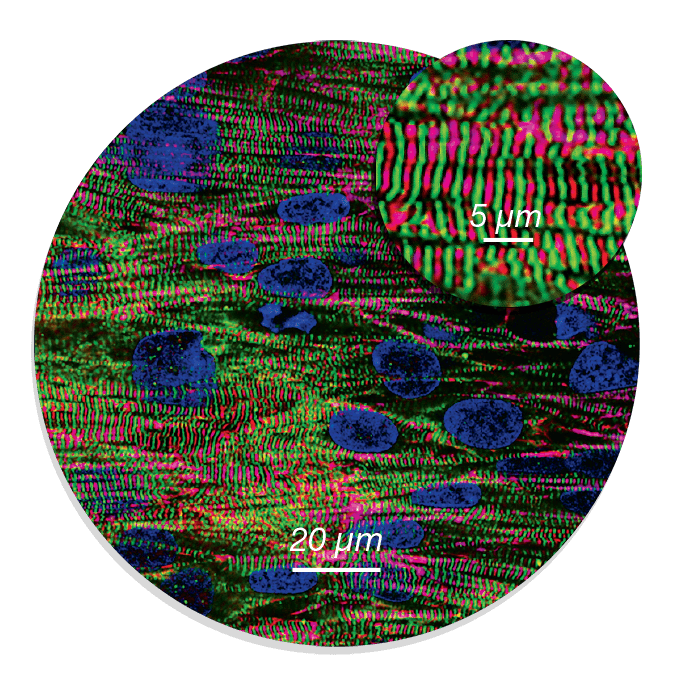
2D Heart Model
Structurally Matured, Aligned Cardiac Microtissues
Curi Bio's 2D heart model utilizes microtissues built by structurally maturing human iPSC-derived cardiomyocytes in biomimetic NanoSurface™ Plates. Structurally mature cardiac microtissues can be engineered from many cell lines – including commercial lines and disease lines – and can be used with a wide range of assay formats and screening workflows.
iPSC-derived cardiac tissues plated on NanoSurface technology are more structurally and functionally mature than standard iPSC-derived cardiomyocyte models from the same cell source, exhibiting:
Polarized expression of gap junction proteins, such as Cx43.
Anisotropic cell shape and aspect ratio, striated sarcomeres, and cytoskeletal alignment.
Enhanced electrophysiological performance, including faster longitudinal conduction velocity, lower resting membrane potential, and longer action potential durations.
Learn more about NanoSurface technology:

Curi Bio's 2D Heart Model Provides
- Enhanced Reproducibility
- High-throughput Screening
- Predictive Drug Responses
- MEA-based Electrophysiology
- Diverse Endpoints

Structurally Mature Human iPSC-Derived Cardiomyocytes

Standard iPSC-CM Cardiac Model

NanoSurface iPSC-CM Cardiac Model
Application Examples
NanoSurface Plates Enhance the Structural Development of Cells and Tissues, Improving the Physiological Relevance of Cell-based Assays
Learn more about Curi Bio’s NanoSurface Plates and how they can benefit your lab:

Curi Bio's 384-well NanoSurface Plate