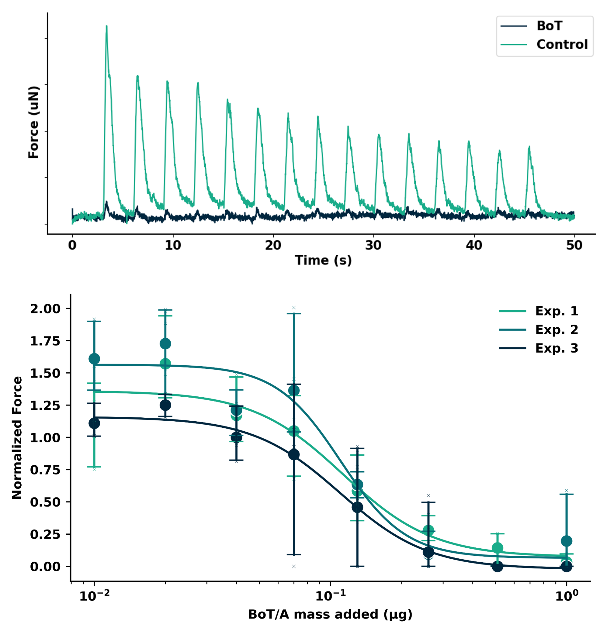
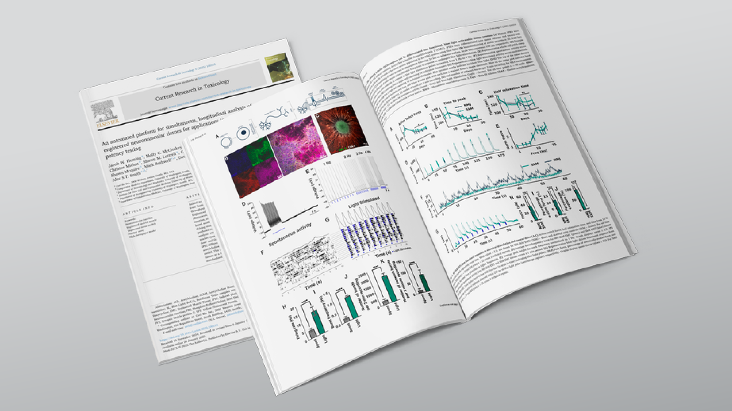
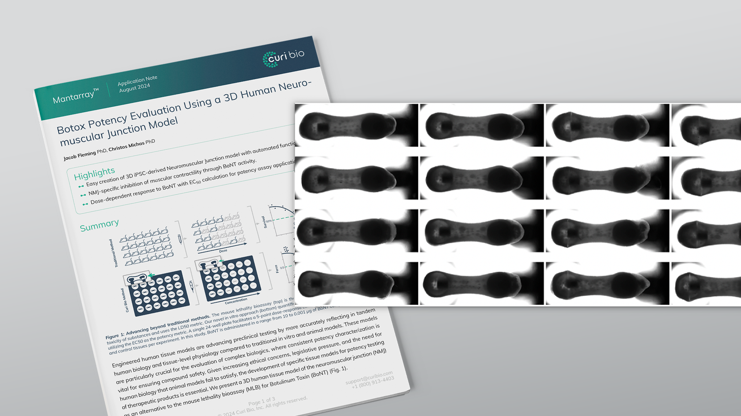
A Novel Human 3D Neuromuscular Junction Model for Drug Discovery
A New Approach to Potency Testing with Scalable Creation and Reliable Results
Histological Characterization of NMJs
Preclinical models often fail to predict how compounds will perform in human clinical trials, leading to a high rate of failure. Current animal models frequently don't accurately reflect human physiology, and many complex biologics are not suitable for these models.
Existing 3D models of the neuromuscular junction (NMJ) have also fallen short, lacking the reliability and high-throughput capabilities needed for effective testing.
The Curi Bio team has designed, built, and tested a functional human 3D model of the neuromuscular junction using our established Mantarray 3D engineered tissue platform. This model offers a solution by incorporating the extracellular matrix (ECM) and cell-to-cell interactions, which significantly improves how well in vitro tests replicate real-world in vivo conditions. This allows for direct assays of disease phenotypes or toxin action, making them perfect for preclinical testing or potency assays. To demonstrate the utility of this model, we show it has a controllable functional output and can be used to study the effects of neurotoxins.
Curi Bio has made the creation of these 3D NMJ models simple and scalable. The process uses iPSC-derived motor neurons and skeletal muscle cells with a high success rate of NMJ creation (over 95%).
In-situ neurosphere creation, which eliminates the need for direct spheroid handling.
Blue light sensitive neurons that allow for specific activation of motor neurons independently of the skeletal muscle.
iPSC derived motor neurons and skeletal muscle.
High success rate of NMJ creation (>95%).
Curi Bio has histological proof of NMJs, which is essential for many disease modeling applications.
Co-cultures exhibited elevated spontaneous contractions compared to single-cultured skeletal muscle tissues. When activated with blue light pulses, the skeletal muscle in the co-culture showed synchronized contractions within 100 milliseconds of the applied light pulses.
Curi Bio demonstrated a dose-dependent loss of NMJ function in the presence of Botulinum toxin (BoT). The tissues showed sensitivity to BoT synapse blockade, with function reduced to 0.03% after four hours of treatment with 5 µg/mL of BoT. This effect was specific to the neuronal component, as the skeletal muscle itself remained functionally competent. Curi Bio also generated a dose-response curve, determining an EC50 value of 0.114 µg after 24 hours of exposure.
This platform serves as a powerful new tool for preclinical drug discovery and a potential solution for potency testing of biological products such as Botulinum neurotoxins.
Easy, rapid casting.
Over 95% casting success rate.
Label-free tracking of 24 tissues.
Well-based electrical stimulation of 24 tissues.
Automated metric extraction.
Clinically relevant protocols.
With the Mantarray platform, you can bypass the limitations of traditional models and get more accurate, reliable results.
If you are interested in learning more about how Curi Bio’s NMJ model can accelerate your research, please contact us to arrange a conversation with our team of experts.