
DM1 Still Has No Approved Disease-Modifying Treatments. Here's What 3D Human Muscle Tissue Reveals.
September 15th is International Myotonic Dystrophy Awareness Day, a day when researchers, clinicians, and patient advocates align around a disease that affects an estimated 1 in 8,000 people worldwide. Despite decades of scientific progress, Myotonic Dystrophy Type 1 (DM1) remains without a single approved disease-modifying treatment.
DM1 is the most common form of adult-onset muscular dystrophy. It causes progressive muscle wasting, difficulty relaxing muscles after contraction (a hallmark symptom called myotonia), fatigue, and in many cases serious cardiac complications. Families living with DM1 know its weight intimately. The research community has characterized its genetic cause in molecular detail. To date, no disease-modifying therapy has reached approval, and patients rely on symptomatic management alone.
Why Preclinical Models Have Struggled to Translate
For years, the standard tools for studying DM1 have been two-dimensional cell cultures and rodent models. Each captures part of the disease, and each leaves out features that matter for measuring therapeutic benefit. 2D cultures do not generate contractile force. They cannot replicate the structural organization of muscle, and they cannot reproduce the sustained contractions that define myotonia. Rodent models have driven most of what the field understands about CUG repeat toxicity and MBNL sequestration, but they carry three constraints worth naming:
Limited somatic repeat instability. Human DM1 muscle accumulates expansion-biased mosaicism into the thousands of repeats, and progression appears to track that expansion. Rodent models expand modestly at best, which limits their usefulness for testing therapies whose benefit depends on slowing or reversing instability.
Compressed disease timescale. DM1 in patients unfolds over decades through cumulative regeneration failure and fibro-fatty replacement. Rodent models tend to present either a largely static phenotype or a severe early-onset one, so neither reproduces the slow degenerative arc.
Divergent splicing landscape. A meaningful fraction of human MBNL-dependent splice events has no direct rodent counterpart, so a splicing signature validated in rodents may not correspond to the signature that matters in patients.
Taken together, these constraints help explain a pattern the field has observed: preclinical models have predicted molecular pharmacology reasonably well, while questions about functional recovery in human muscle have remained harder to answer before the clinic.
To address that gap, Curi Bio built a complementary human model.
A Human Model That Contracts
Working with patient-derived induced pluripotent stem cells (iPSCs) carrying defined DM1 repeat expansion lengths, Curi Bio scientists developed an assay-ready platform of 3D Engineered Muscle Tissues (EMTs) that contract and generate measurable force. The platform pairs these patient lines with an isogenic CRISPR-corrected control, a genetically matched line in which the disease-causing CTG repeats have been removed. Pairing the lines isolates the contribution of the repeat expansion from the donor genetic background.
Three Key Findings
Curi Bio researchers evaluated DM1 tissues against healthy controls across three dimensions of disease biology.
Finding 1: Contractile Force Tracks Repeat Expansion Length
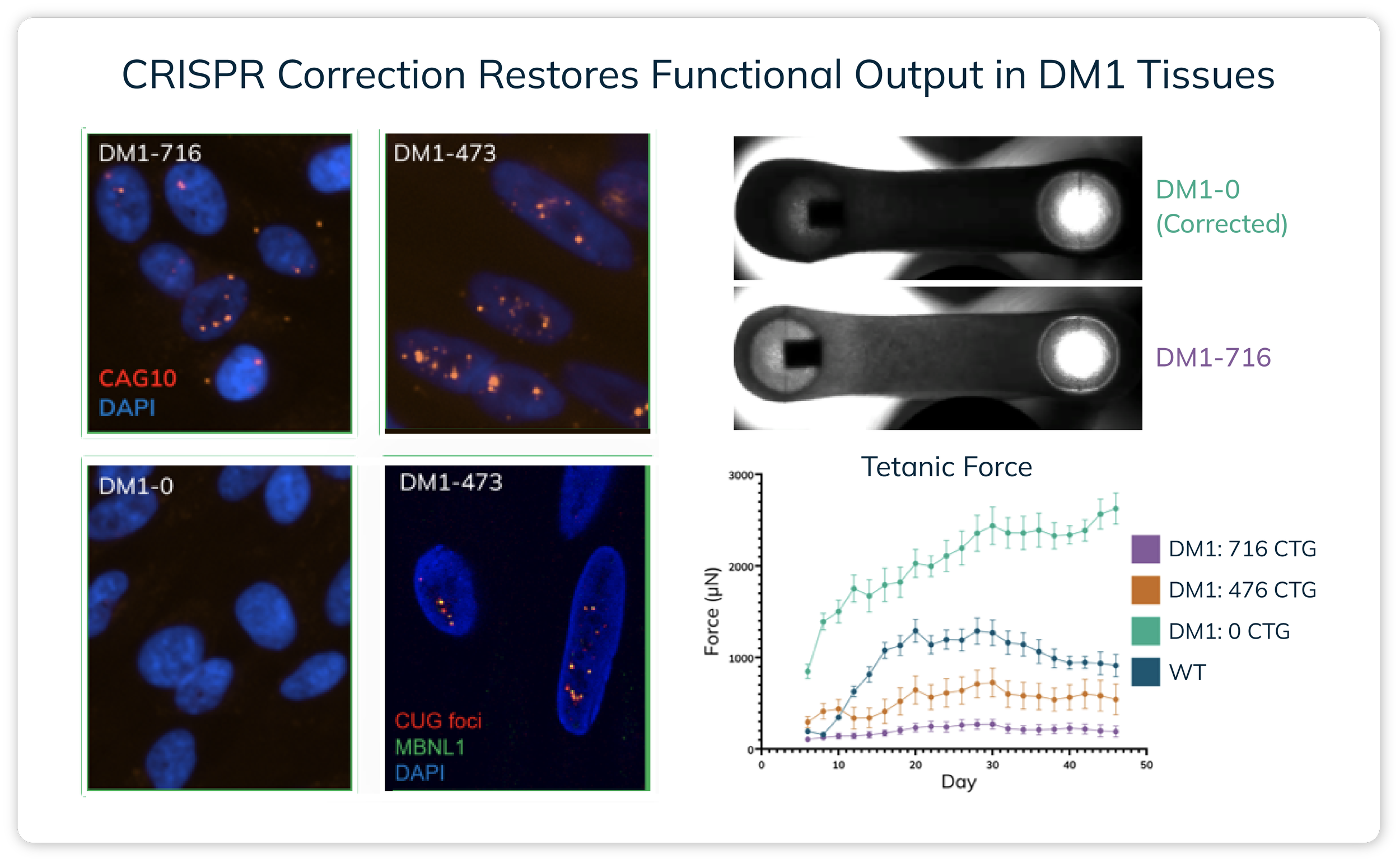
Over 46 days of longitudinal culture, tissues carrying more severe DM1 repeat expansions generated less contractile force, falling below one-third the output of the CRISPR-corrected control at peak. The Mantarray™ platform measures this force non-invasively across the full culture period. The result is consistent with the clinical relationship between repeat length and disease severity, in which longer expansions are associated with earlier onset and greater motor impairment.
Finding 2: Gene Correction Restores Function
In the isogenic corrected line, contractile force returned to healthy control levels, and the toxic nuclear RNA aggregates central to DM1 pathology were largely eliminated. This establishes a functional benchmark for rescue, a measurable reference point against which therapeutic candidates can be tested.
Finding 3: Calcium Kinetics Reveal Delayed Relaxation in DM1 Tissues
Using the Nautilai™ platform for calcium imaging, disease tissues showed prolonged calcium influx and delayed relaxation relative to controls. These kinetics are consistent with the impaired relaxation that characterizes DM1 muscle, and the readouts are quantifiable, reproducible, and specific to the disease lines.
What This Means for Drug Discovery
Contractile force. RNA foci clearance. Calcium transient kinetics. Together, these three readouts form a functional fingerprint for DM1 in human tissue, one that 2D cultures and rodent models do not provide.
For researchers developing antisense oligonucleotides, small molecule splicing modulators, or gene therapies, the platform measures whether a candidate restores human muscle function before a program commits to clinical trials. It connects molecular mechanisms to functional recovery in the tissue that matters.
Our Commitment to the Neuromuscular Field
At Curi Bio, we believe preclinical models should earn their place in drug discovery programs. For DM1, and for every neuromuscular condition where patients are still waiting, that means models that behave like human tissue, generate data that mirrors clinical observation, and give research teams a functional basis for advancing a candidate.
On International Myotonic Dystrophy Awareness Day, we are reminded that the work is not abstract. The patients and families living with DM1 deserve better tools in the hands of the researchers working to help them. We are proud to be part of that effort.
Download the Application Note
Explore the full dataset, methodology, and results in our application note:
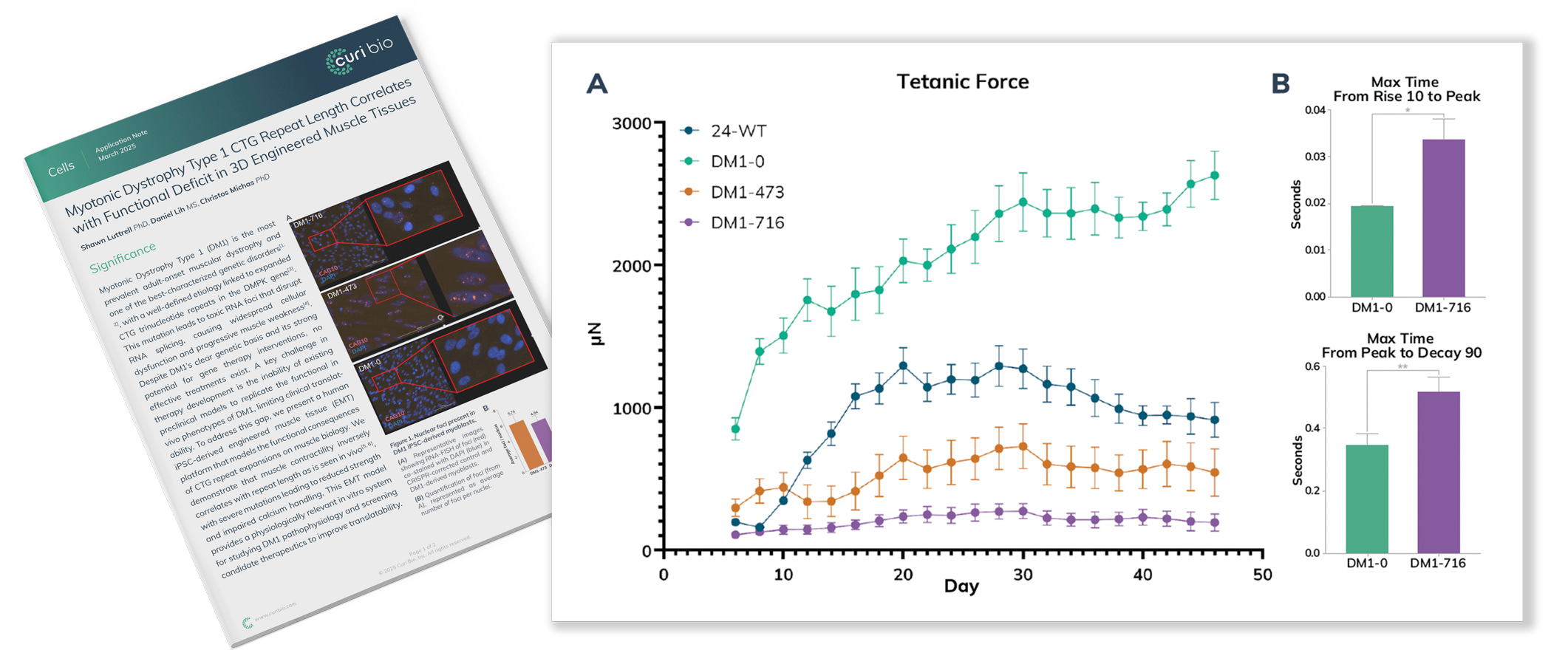
"Myotonic Dystrophy Type 1 CTG Repeat Length Correlates with Functional Deficit in 3D Engineered Muscle Tissues"
Speak with A Team Member
Interested in integrating 3D engineered muscle tissues into your preclinical pipeline? Contact a Curi Bio team member to learn how our custom research services and human-relevant functional readouts can help de-risk your DM1 discovery program.