Cart 0
Cart 0
 
MYBPC3 Hypertrophic Cardiomyopathy Isogenic Pair Cell Line

Isogenic MYBPC3 Hypertrophic Cardiomyopathy Model

Cryopreserved, Assay-ready Cardiomyocytes for Advanced Cardiac Disease Research

iPSC-Cardiomyocyte (iCM) Line Genetic Mutation Clinical Disease Price
CB-iCM-MYBPC3-e6-het
(Mutant)
Frame shift mutation in exon 6. Hypertrophic Cardiomyopathy $2500
5 million cells
CB-iCM-MYBPC3-e6-corr
(Isogenic Control)
CRISPR-corrected mutation. $2500
5 million cells
Request a Quote to Order Speak with an Expert
 
curi-bio-gradient.png
 

Definitive Model for Hypertrophic Cardiomyopathy Research

Hypertrophic Cardiomyopathy (HCM) is the most common genetic heart disease, affecting an estimated 1 in 500 individuals worldwide. Mutations in the MYBPC3 gene are the single most common cause of HCM*, accounting for up to 50% of all familial cases.

Accelerate your drug discovery and mechanistic studies for HCM with the MYBPC3e6 Isogenic Pair—the industry's first commercially available hypertrophic cardiomyopathy isogenic pair cell line. This validated model provides an unmatched platform to precisely study the disease phenotype and evaluate therapeutic candidates.

*Garcia-Hernandez et al., Can J Cardiol, 2024

What is the MYBPC3e6 Isogenic Pair?

This groundbreaking pair consists of an induced Pluripotent Stem Cell (iPSC) line carrying a pathogenic mutation and its genetically CRISPR-corrected control.

The mutant line was generated from peripheral blood mononuclear cells of a male Caucasian patient diagnosed with hypertrophic cardiomyopathy. The isogenic control provides the critical, genetically identical wild-type reference to isolate and study the specific effects of the MYBPC3 mutation.


Key Disease Phenotypes in Engineered Heart Tissues (EHTs)

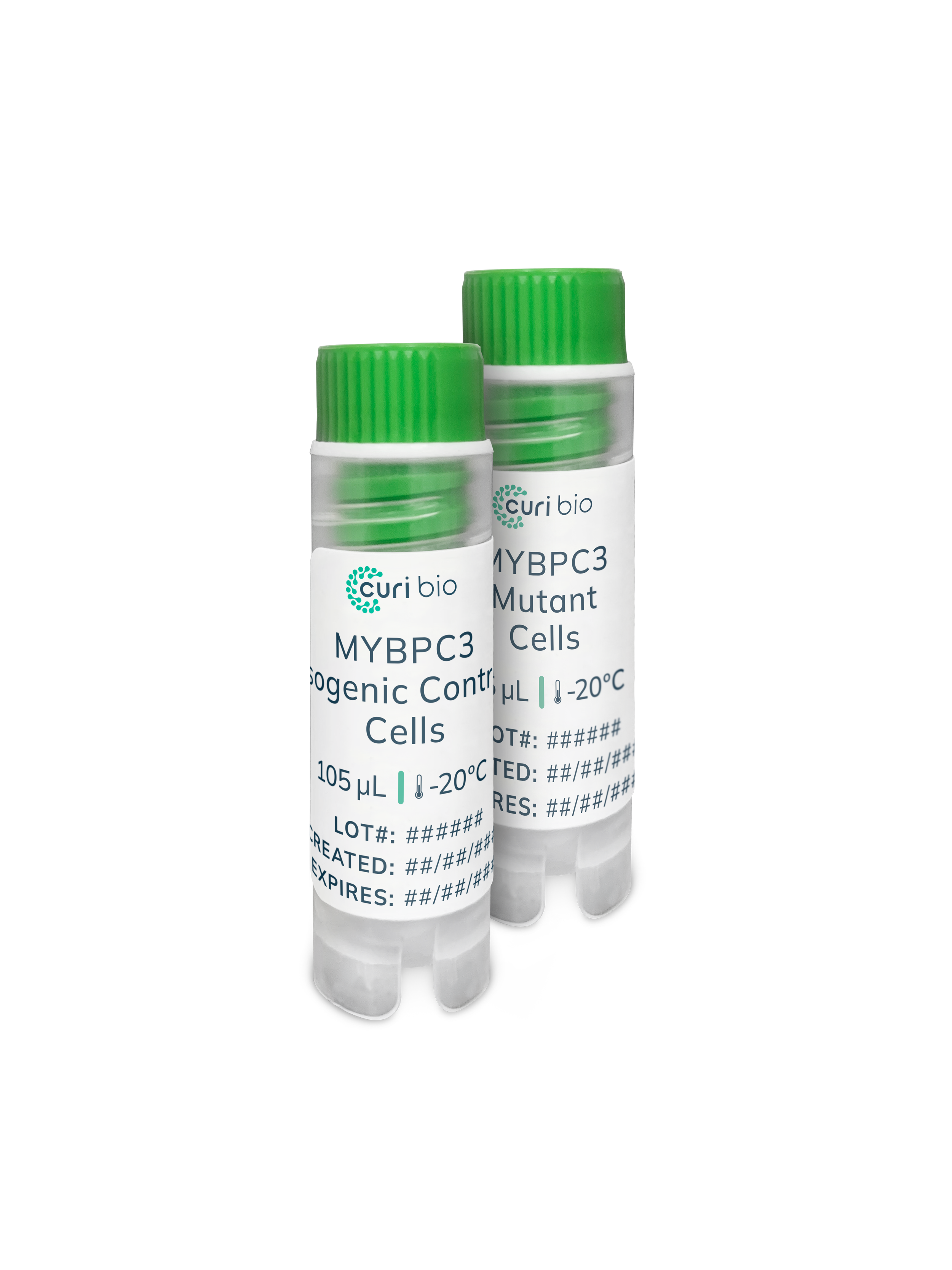
Our MYBPC3e6 cardiomyocytes, when modeled as Engineered Heart Tissues (EHTs) on Curi Bio’s Mantarray™ and Nautilai™ platforms, reliably display hallmark HCM phenotypes:

1. Hypercontractility & Calcium Hypersensitivity

The MYBPC3e6 mutant tissues exhibit hypercontractility during EHT maturation when continuously electrically paced. They also show a heightened inotropic response to extrinsic calcium, suggesting an underlying defect in calcium handling or myofilament sensitivity that drives excessive force generation.

  • Twitch Force Comparison: At high calcium concentrations (4mM and 6mM), the mutant line produces a significantly higher twitch force (up to ∼850% change) compared to its isogenic control (∼450% change).

2. Impaired Relaxation Kinetics

HCM is fundamentally a diastolic dysfunction disorder. The MYBPC3e6 model demonstrates significantly altered relaxation kinetics that are rescued by the myosin inhibitor Mavacamten, a compound clinically used for HCM treatment.

  • Relaxation Time: The MYBPC3e6 tissues show a statistically significant increase in the time from Peak to Relaxation 50 compared to the Isogenic Control, indicating slower relaxation.

  • Pharmacological Rescue: Treatment with 0.1 μM of Mavacamten reverses the slowed relaxation kinetics, restoring the relaxation time to levels statistically similar to the isogenic control.

3. Failure to Maintain Pacing at Low Calcium

Under stress testing, the MYBPC3e6 tissues fail to follow high-frequency electrical pacing, a finding that reflects the impaired chronotropic reserve often observed in HCM patients. This failure is most pronounced at low calcium levels, demonstrating the model's sensitivity to metabolic stress.

  • Stress Test Failure: At a stimulation frequency of 3.5 Hz, nearly 90% of the mutant tissues fail to follow pacing in 1 mM calcium conditions, whereas the isogenic control remains at 100% following pacing.

Advanced Mechanistic Insights

Leverage the MYBPC3e6 Isogenic Pair to explore deeper molecular mechanisms and test novel drug candidates.

4. Reduced Response to Myosin Activation

The mutant line displays an attenuated response to the myosin activator EMD57033, providing a unique screening window for compounds targeting the contractile machinery.

  • EMD57033 Treatment: Following treatment with 10 μM EMD57033, the increase in twitch force is significantly lower in the MYBPC3e6 tissues compared to the Isogenic Control.

5. Altered Electrophysiology in Hypercalcemia

Action potential (AP) measurements in hypercalcemic media (4 mM Ca2+) reveal key electrophysiological differences, measured using Curi Bio’s Nautilai™ platform.

  • Fast Upstroke Time: At all pacing frequencies, the MYBPC3e6 tissues exhibit a faster Time from Rise 10 to Peak (AP upstroke time) compared to the isogenic control.

  • Delayed Repolarization: At 3 Hz and 4 Hz pacing, the MYBPC3e6 tissues show a significantly slower Time from Peak to Decay 50 (repolarization time).


Begin Your HCM Research Today

With the MYBPC3e6 Isogenic Pair, you gain:

  • Genetic Fidelity: An isogenic control created by CRISPR/Cas9 for high-confidence results.

  • Physiological Relevance: Functional validation using cutting-edge Engineered Heart Tissues (EHTs) and Curi Bio's proprietary platforms.

  • Translational Power: Phenotypes, including hypercontractility and impaired relaxation, that are clinically relevant and responsive to known therapeutics like Mavacamten.

Stop wasting time and resources. Request a quote or speak to a specialist to integrate the MYBPC3e6 Isogenic Pair into your research workflow.


More MYBPC3 Resources

Go To App Notes Page